Diagnostics and dental X-rays
Dental X-rays – Pantomographic images and tomography Wrocław MedicaDent
A precise diagnosis is absolutely essential for effective and safe dental treatment. At MedicaDent in Wrocław, we understand that it is impossible to plan treatment at the highest level without an accurate image of the interior of the tissues. That is why our practice is based on modern digital X-ray diagnostics, including advanced 3D dental tomography. Thanks to this, we can provide our patients with accurate diagnoses and, above all, maximum safety.
A full range of X-ray imaging examinations at MedicaDent Wroclaw
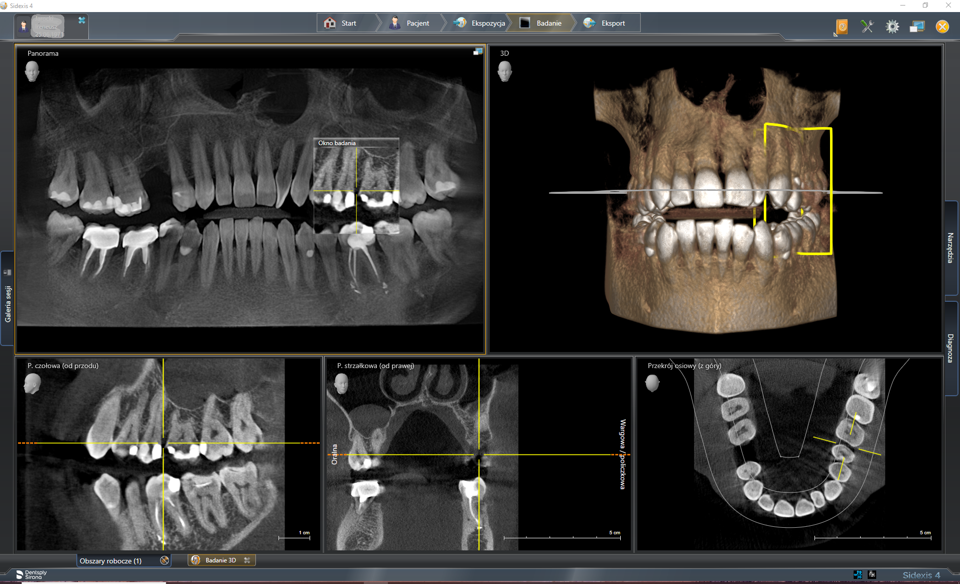
Our diagnostic laboratory is equipped with state-of-the-art technology from the German company SIRONA, including the ORTHOPOS XG 3D CBCT Tomography system. This enables us to perform all the imaging tests required in modern dentistry. Each test is individually tailored to the patient’s clinical needs.
A panoramic (pantomographic) image provides a comprehensive view of the teeth.
Often referred to as a panoramic dental image, a pantomographic image is a basic overview examination. This single image provides a complete picture of all the teeth (both visible and retained), the maxilla and mandible, the temporomandibular joints, and the maxillary sinuses.
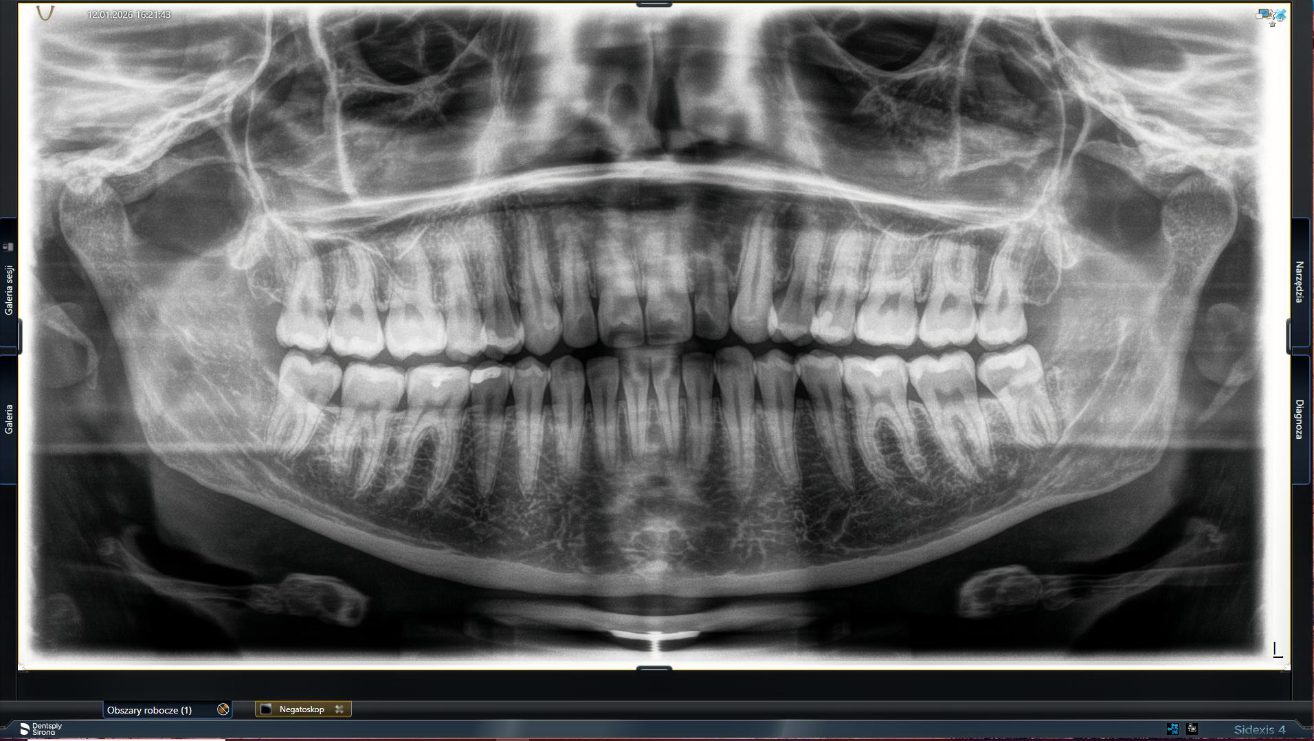
It is an ideal tool for assessing the overall health of the oral cavity, and is the starting point for planning most comprehensive therapies. If you are looking for a place to have a dental panoramic X-ray in Wrocław, our clinic offers this examination on site.
3D computed tomography (CBCT) – the highest precision in implantology and surgery
We use dental tomography (CBCT) when a two-dimensional image is not enough. This is the most advanced imaging test, providing an extremely detailed, three-dimensional model of bone structures. It is indispensable for:
- planning implant treatment, allowing for precise measurement of bone height and width.
- before complex surgical procedures, e.g. the removal of impacted wisdom teeth.
- advanced endodontics for diagnosing unusual root canal anatomy.
A cephalometric X-ray is the key to precise orthodontic treatment planning.
This specialised X-ray image shows a lateral projection of the skull. It is an essential diagnostic tool in orthodontics. It allows orthodontists to take precise measurements, assess the mutual relations between facial bone structures, diagnose malocclusion, and plan effective treatment.
Spot dental X-rays (adjacent) – accurate diagnosis of individual teeth
When the problem concerns one or more adjacent teeth, the best solution is to take a small spot X-ray. This provides an image with the highest resolution and perfect detail reproduction. This is standard practice when diagnosing caries, assessing the tightness of fillings, or checking the effectiveness of root canal treatment. At MedicaDent, we perform dental X-rays in Wrocław without a referral from your GP if the clinical situation requires it.
Diagnosics – frequently aked questions
Modern and safe equipment – why choose MedicaDent?
Patient safety is our priority. We have invested in equipment that combines the highest image quality with minimal exposure to the body.
- Modern digital technology: At our clinic, we use the ORTHOPOS 3D CEPH pantomograph with TOMOGTAF CBCT from SIRONA, among other things
- Highest image quality: Advanced Galileos software and excellent optics guarantee images with extraordinary detail, enabling more accurate diagnoses.
- Availability: We perform dental tomography, cephalometric imaging and dental X-rays in Wrocław, not only for our own patients, but also on request from other dental clinics.
Don’t delay your diagnosis. Contact us to arrange an examination or to ask our specialists which type of imaging would be most suitable for you.